Retina Case #1
Authors: Carolin Aizouki (1), Dr. Mark Seamone (1,2)
Affiliations: (1) University of Alberta (2) Alberta Retina Consultants
ID: 62M; diabetic retinopathy check (no previous dilated fundus exam).
Past Ocular History:
None
Ocular gtts: None
Relevant Medical History: Type 2 diabetes mellitus (HbA1c - 9.3%), HTN (systolics average around 150s)
Medications: Ramipril, insulin, metformin


-
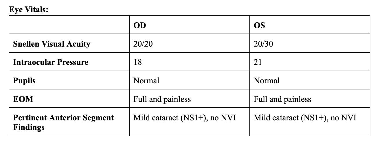
IVFA at 10 seconds
Choroidal phase (early arterial)
Key features include:
Absence of dye in retinal vessels
Faint choroidal flush with patchy appearance of choroid. May be delayed in the context of retinal ischemia.
-
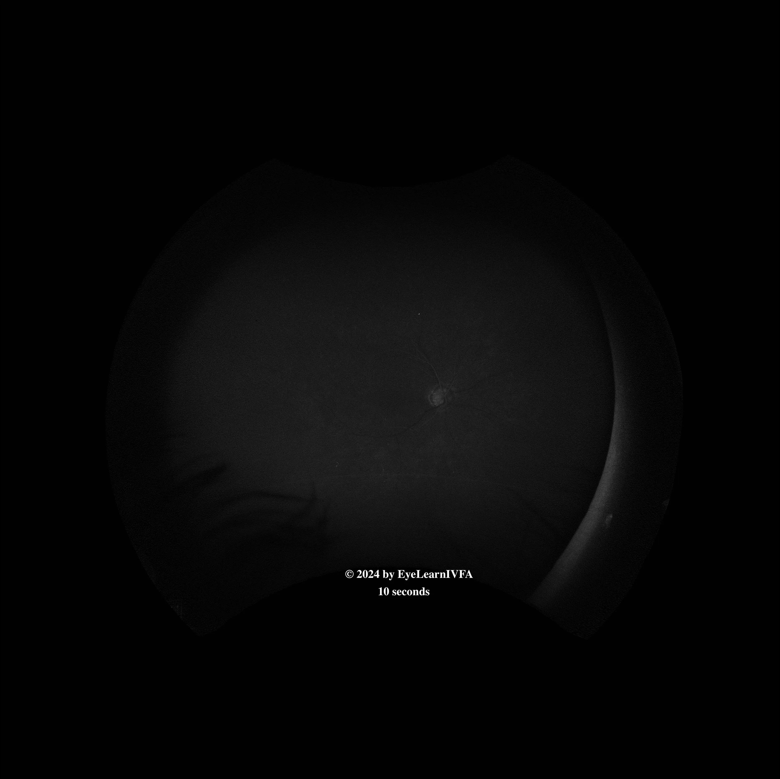
IVFA at 17 seconds
Arterial phase
Key features include:
Several hyperfluorescent dots concentrated around the vasculature representing microaneurysms (1)
-
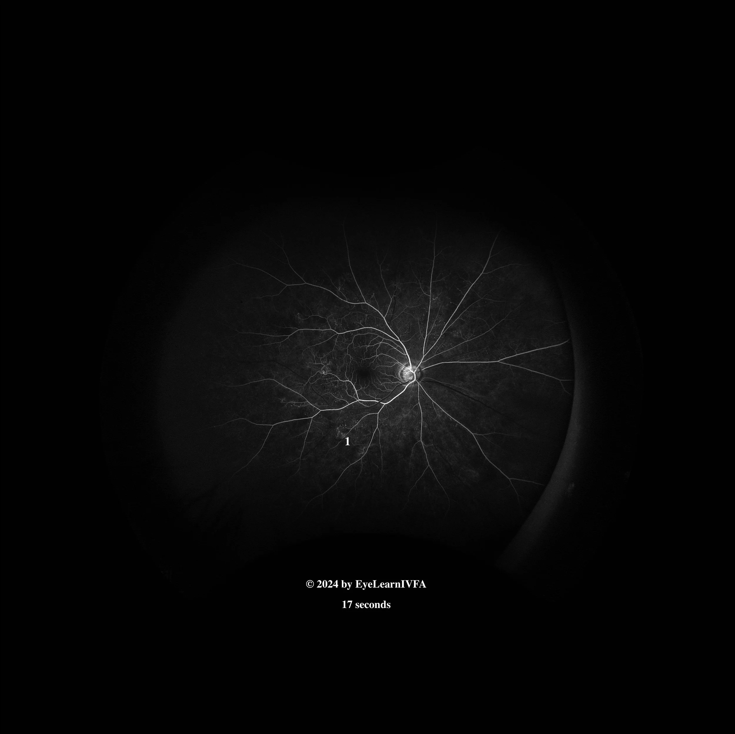
IVFA at 20 seconds
Early arteriovenous phase with laminar flow.
Key features include:
Persistent focal spots of hyperfluorescence secondary to microaneurysms (1).
Faint areas of hyperfluorescence with irregular borders at the edges of the posterior pole secondary to neovascularization elsewhere (NVE). (1)
Hypofluorescence in the supra-temporal region and inferonasal region from non-perfusion secondary to capillary drop-out (2).
-
IVFA at 30 seconds
Late arteriovenous phase
Key features include:
Increasing intensity and size of hyperfluorescence characterized by indistinct borders at areas of NVE secondary to leakage (1).
More apparent areas of capillary drop-out/non-perfusion (2).
IRMAs (Intraretinal microvascular abnormalities), which are dilated intraretinal channels that do not leak (no surrounding hyperfluorescence with obscure/indistinct borders) (3).
No obvious hyperfluorescence around the optic disc to indicate neovascularization of the disc (NVD)
-
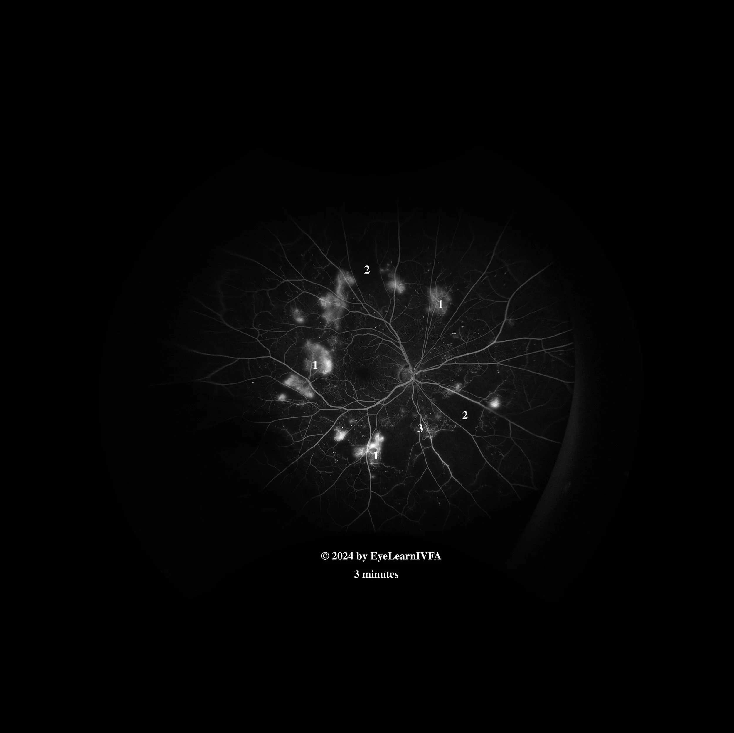
IVFA at 3 minutes
Late phase
Key features include:
Increasing intensity and size of hyperfluorescence characterized by indistinct borders at areas of NVE secondary to leakage. Notice how the leakage extends beyond vessel borders (1).
Continued presence of regions of capillary non-perfusion (2) and IRMAs (3) as previously described.
-
This IVFA is showing a pattern of proliferative diabetic retinopathy, highlighting areas of hypofluorescence due to retinal ischemia and areas of hyperfluorescence secondary to NVE. Many features of diabetic retinopathy in general including IRMAs and MAs are also seen in this IVFA.
-
Causes of retinal ischemia with neovascularization:
Proliferative diabetic retinopathy
Sickle cell retinopathy
Eales’ disease
FEVR
Radiation retinopathy (although patient does not have a history)
Retinal vein occlusions (usually unilateral, would by atypical bilaterally)
Inflammatory diseases with retinal ischemia (retinal vasculitis, Birdshot chorioretinopathy, sarcoidosis, susac syndrome, MS)
Hereditary causes - Incontinentia Pigmenti, Retinitis Pigmentosa
-
In an individual with a longstanding history of uncontrolled diabetes and an elevated HbA1c, presenting without a history of regular dilated exams, IVFA becomes a powerful tool to search for extent and evidence of neovascularization, some of which may be obvious on dilated fundus exams and some of which may be subclinical. As well, IVFA helps to characterize the specific regions of capillary dropout and retinal ischemia. This is imperative for guiding focal laser treatment, initiation and continuation of anti-VEGF treatment and the need for pan-retinal photocoagulation.
-
Chaudhary S, Zaveri J, Becker N. Proliferative diabetic retinopathy (PDR). Disease-a-Month. 2021 May;67(5):101140. doi:10.1016/j.disamonth.2021.101140
Kollias AN, Ulbig MW. Diabetic Retinopathy. Deutsches Aerzteblatt Online. 2010 Feb 5;107(5). doi:10.3238/arztebl.2010.0075
Kour V, Swain J, Singh J, Singh H, Kour H. A Review on Diabetic Retinopathy. Current Diabetes Reviews. 2024 Jul;20(6). doi: 10.2174/0115733998253672231011161400