Anatomy Overview
What are the layers of the choroid and their associated characteristics?
Choroid: the posterior segment of the uvea, providing blood supply to the outer retina.
Composed of 5 layers:
Bruch's membrane: Separates the retinal pigment epithelium from the choroid.
Choriocapillaris: Dense network of fenestrated capillaries, creating high oncotic pressure that drives fluid from retina to choroid.
Sattler's layer: Medium-sized arteries and arterioles.
Haller's layer: Large blood vessels.
Suprachoroid: Transition zone between choroid and sclera.

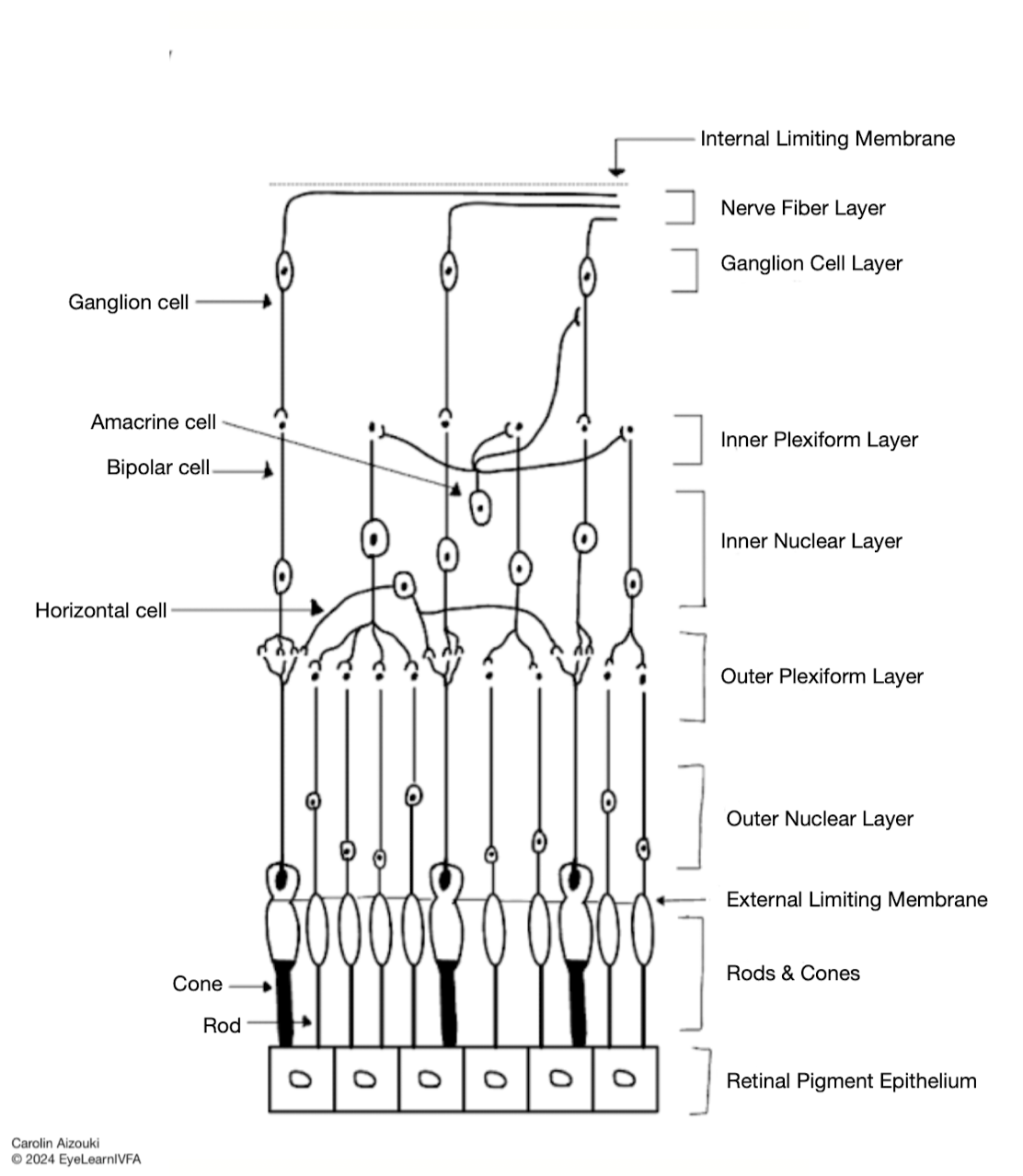
What are the layers of the retina and the cells within them?
Innermost
Inner Limiting Membrane (ILM): Separates the retina from the vitreous.
Nerve Fiber Layer: Composed of the axons of retinal ganglion cells. This layer’s basal lamina is the ILM.
Ganglion Cell Layer: Cell bodies of ganglion cells.
Inner Plexiform Layer: Bipolar cell axons synapse with ganglion cells. It also includes synapses from amacrine cell dendrites to modulate electrical conduction between bipolar and ganglion cells.
Inner Nuclear Layer: Cell bodies of bipolar cells, horizontal cells, and amacrine cells.
Bipolar cells transmit synaptic inputs from photoreceptor cells to ganglion cells.
Horizontal cells modulate signals from rods and cones.
Outer Plexiform Layer: Axons of photoreceptor cells synapse with cells within the inner nuclear layer.
Outer Nuclear Layer: Cell bodies of both rods and cones.
External Limiting Membrane: Separates cell bodies of rods and cones from their inner and outer segments.
Outermost