Practice the Basics: Hyperfluorescence
Pattern #1: Pre-injection Fluorescence
Pre-injection fluorescence: fluorescence in images taken before the fluorescein dye is injected. Two subcategories are autofluorescence and pseudofluoresence.
What is autofluorescence?
Fluorescence prior to dye injection due to the presence of fluorophores such as lipofuscins or porphyrins.
Optic disc drusen can have autofluorescence due to the presence of excessive mitochondria that contain porphyrins.
Retinal astrocytic hamartomas may also show autofluorescence.
What is pseudofluorescence?
May occur due to old filters which allow transmission of light outside of the specified blue and green wavelengths.
May be caused by dehemoglobinized blood - reflects blue light and will present as hyperfluorescence.
Pattern #2: Transmitted Fluorescence or Window Defect
Pattern #3: Leakage
A leak occurs when fluorescein enters the extravascular space.
It begins early in the scan and increases in size and intensity with time. Borders are usually indistinct.


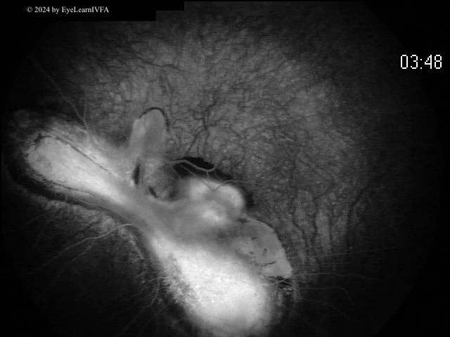
Macular Leakage: Type 2 CNVM
Notice the indistinct borders and the early hyperfluorescence that the increase in both size and brightness throughout the fluorescein angiogram.
Because dye illuminates retinal vessels well, it is quite easy to determine the presence of abnormal retinal vasculature.
Abnormal vasculature can appear in the choroid or retinal layers.
Pattern #4: Pooling
The accumulation of leaked fluorescein within a defined anatomical space.
Once this defined space is filled, the margins remain constant and distinct throughout the study (unlike leakage which continues to grow in size and brightness the scan progresses).
Pattern #5: Staining

Staining happens when fluorescein dye molecules attach to structures.
The margins or amount of fluorescence in staining does not intensify with time and does not go beyond the pathology (unlike leaking).
Staining usually involves later hyperfluorescence; it is usually mild and not bright.
Toxoplasmosis Scar
This is a later phase fluorescein angiogram that shows a large inferior atrophic chorioretinal scar from congenital toxoplasmosis.
Physiologic Staining
Staining can also be physiologic; structures that stain in late phases include the optic disc, Bruch's membrane, and the sclera.
Pattern #5: Neovascularization (A Subpattern of Leakage)
Pooling due to Central Serous Retinopathy
Notice how, in comparison to leakage, the area remains roughly the same size between early and late stages, with a subtle increase in fluorescence intensity.

Now:
-
Tortuosity and dilation: think diabetes or vascular occlusion.
Collaterals and Shunts: classify as arterial, arterio-venous or venous-venous.
Neovascularization: classify it as in the posterior pole or periphery. Think of diabetes, sickle cell disease, vascular occlusions, retinopathy of prematurity.
Aneurysms: microanuerysms from diabetes or vein occlusion or larger aneurysms like in tumours, Coat's, Leber's and macular telangiectasis.
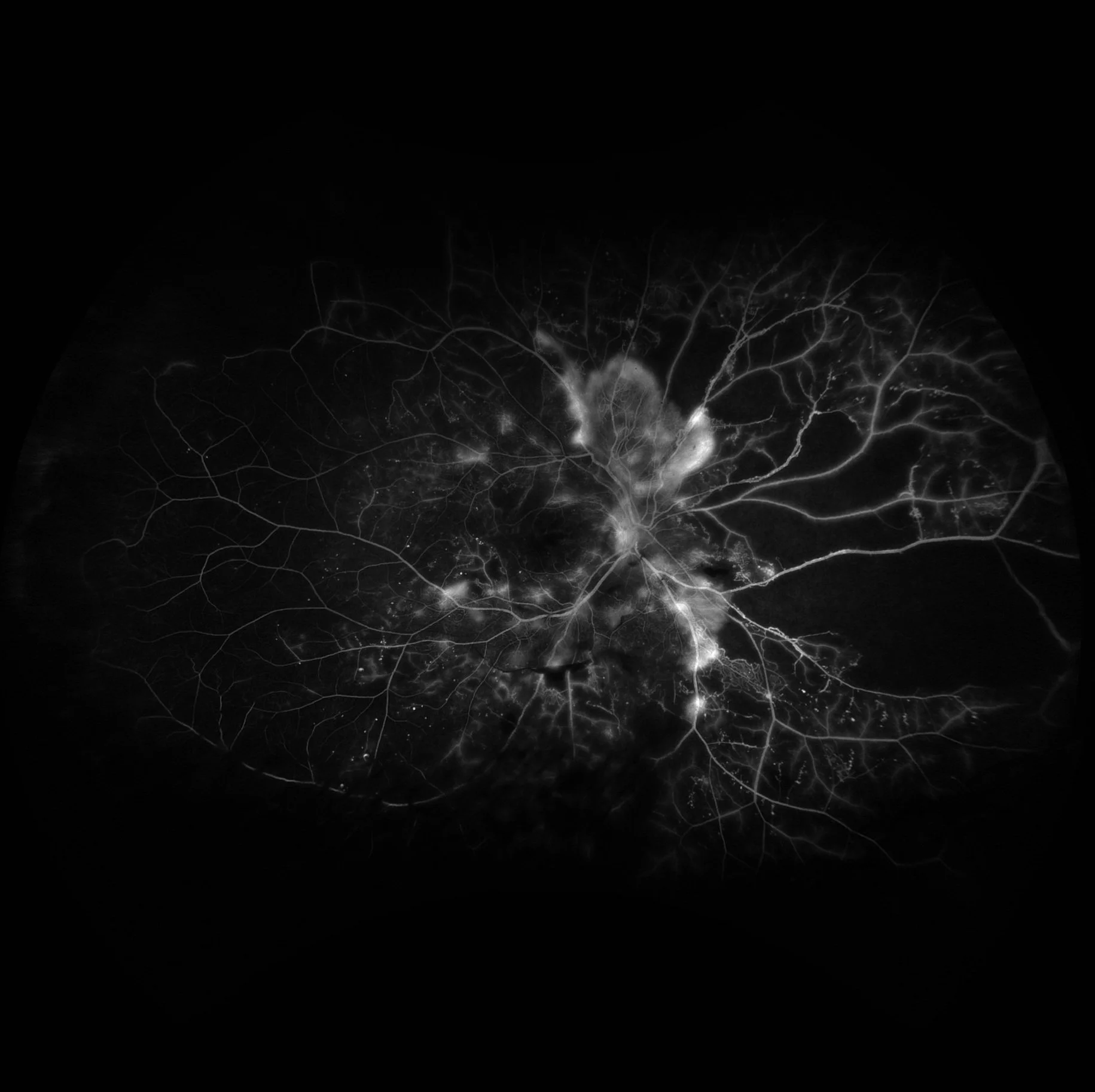

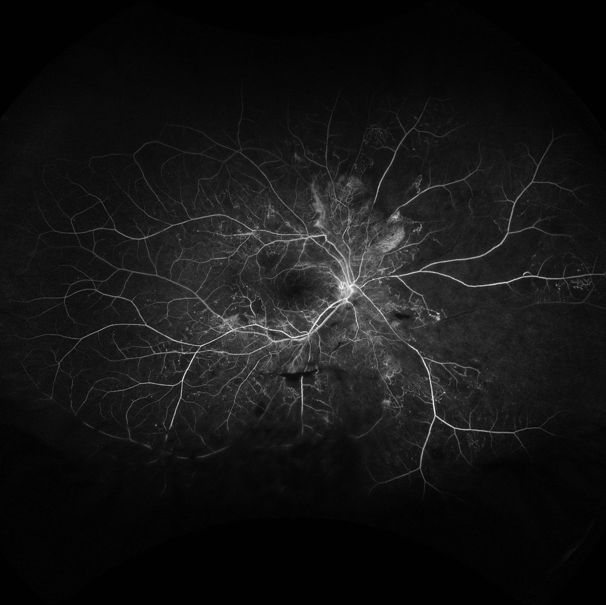
Proliferative Diabetic Retinopathy
This is a severe case of proliferative diabetic retinopathy, with neovascularization at the disc and peri-papillary areas. Abnormal vessel formation, that are friable and leaky, lead to hyperfluorescence with indistinct borders that widens and intensifies as the angiogram progresses.
Notice the extensive occlusive disease and non-perfusion due to capillary drop out in the periphery.
Healthy RPE blocks choroidal fluorescence, if there is a RPE defect there will be hyperfluorescence of underlying choroid layer.
Hyperfluorescence occurs in the same shape as the RPE defect - it does not increase in size or shape with time.
Window defects fade as phases of the scan progress and the dye washes out of choroidal circulation.

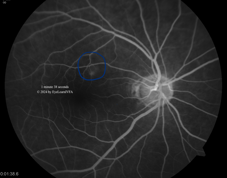
Transmitted Fluorescence Due to Diffuse Drusen
Hard drusen are yellow deposits in Bruch's membrane, made of hyaline material. They appear hyperfluorescent on IVFA due to RPE thinning, with atrophy further revealing choroidal fluorescence. Not all drusen hyperfluoresce, as this depends on RPE thinning and atrophy