Hyperfluorescence Case #5
ID: 69F
Reason for Referral: Central Scotoma NYD
Past Ocular History: None
Past Medical History: Hypertension, Diabetes (HbA1c 6.1%)
Medications: Metformin
Ocular gtts: None
Eye Vitals and Examination:
BCVA 20/60 OD, 20/25 OS
IOP 16 OD, 15 OS
No RAPD, PEARRLA
EOM full
-
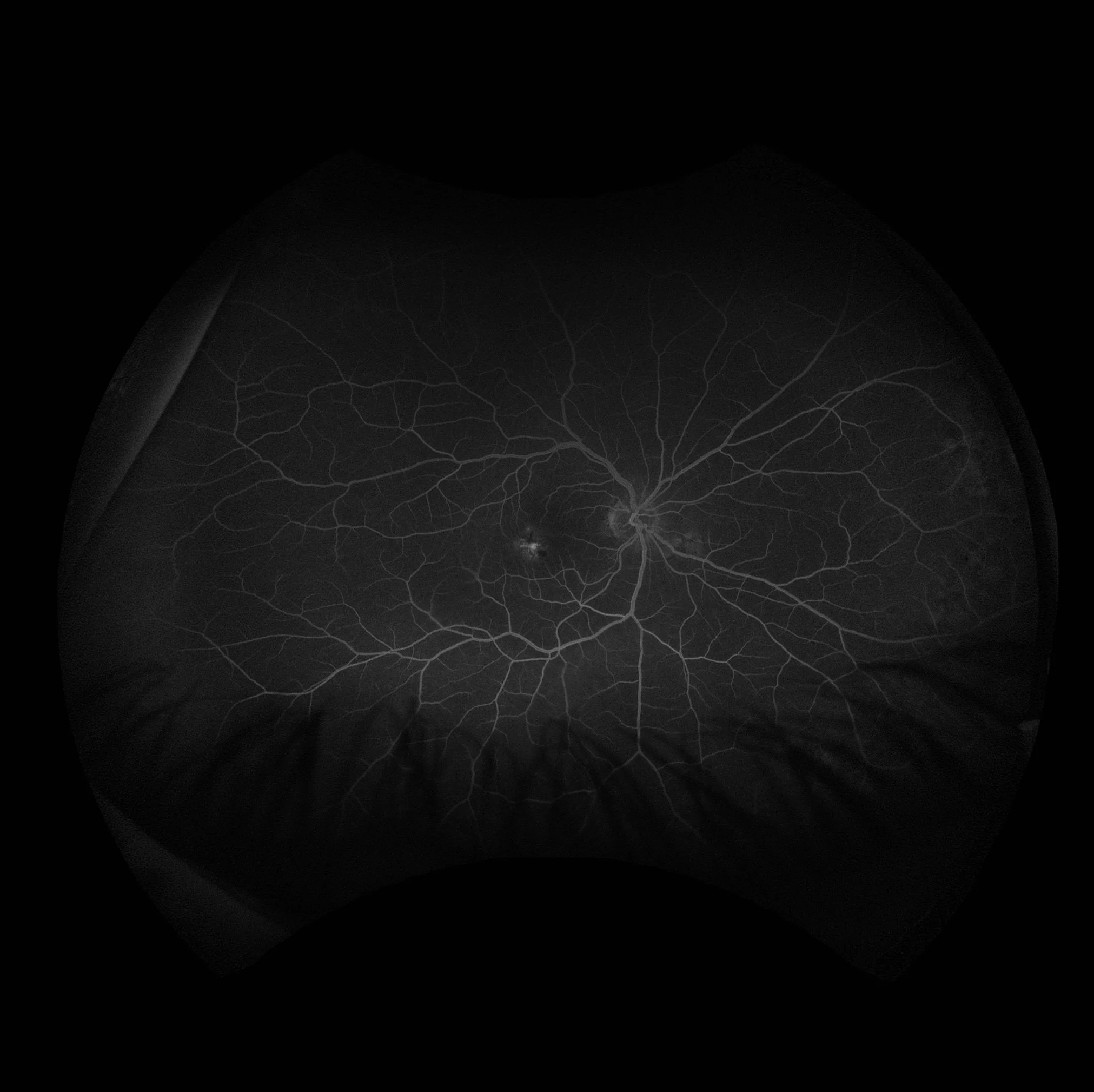
Late Leakage at the Macula
A leak occurs when there is loss of fluorescein into the extracellular space. A full leak begins early in the scan and increases in size and intensity with time. This photo shows late macular leakage and right angled vessels characteristics of macular talengiectasia.
-
Cystoid appearance (inside the macula, aflower petal appearance because of the organization of the outer plexiform layer; outside of the macula, a honey- comb appearance): diabetic retinopathy, telangiectasis, choroidal or retinal inflammation, subretinal neovascularization, subretinal inflammation, choroidal tumours.
Non-cystoid pattern (usually due to small retinal vessel leakage): hypertension, diabetes, or vasculitis.